In the field of neuroscience, it could be said that imaging is everything. Being able to look inside the brain at structure, injury or illness is crucial for medical professionals and researchers to understand how the brain works and what to do when it doesn’t work as it should.
There are constant advances being worked on in the field of brain imaging. One of the most well known is magnetic resonance imaging, or MRI. Scanning a patients brain with an MRI machine can reveal any number of problems from strokes and blood clots, to tumors and neurodegenerative diseases.
MRIs work by harnessing the power of extremely strong magnets along with radio frequency waves to image tissues in the brain. They are extremely complex from an engineering point of view, but they can produce pictures of the brain that stunningly clear. One of the advances in the technology of MRIs is a functional MRI or fMRI. When a patient is in the MRI scanning tube, they are asked to complete a cognitive task like viewing pictures, repeating words or doing simple math. While this is happening, the activity in the brain is being recorded. Scientists can see, in real time, what parts of the brain and which neurons are being activated when certain tasks are completed.
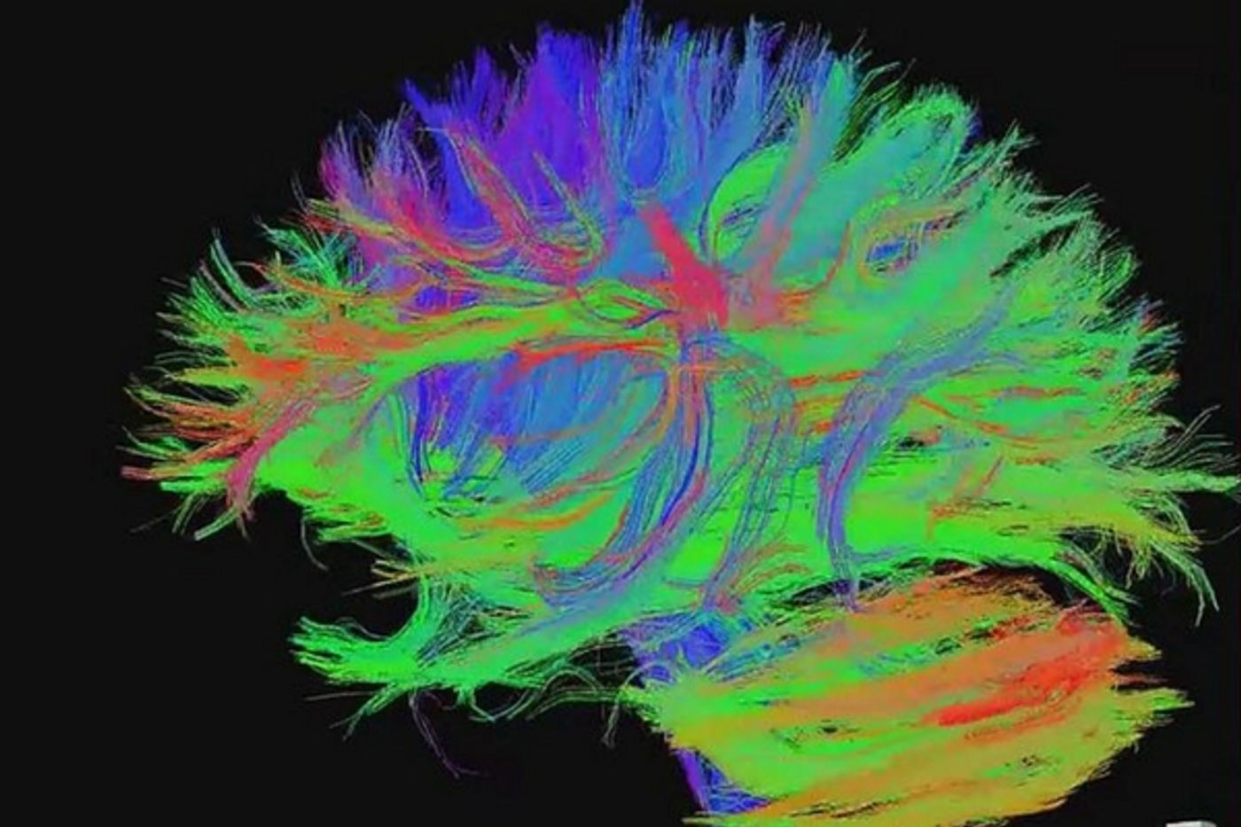
Diffusion weighted MRIs or DW-MRI scans are also important breakthroughs in MRI technology since they can pick up the diffusion of water molecules in the brain. Specifically in a DTI MRI, or diffusion tensor imaging scan, the architecture of white matter in the brain can be seen and these images can yield clues to disease progression and the extent of any brain injury a person has sustained.
Recently, at Cardiff University in Wales, a new scanner was demonstrated for the BBC audience. The machine, which is known as a microstructural MRI scanner, is part of the Cardiff University Brain Research Imaging Centre (CUBRIC) and is one of only three of it’s kind in the world. What it does is map the actual axons in the brain, the fibers that are the wiring in the brain along which neurons send their signals. It would take 50 axons to equal the size of one human hair, so they are incredibly small structures considering the large part they play in keeping the brain working correctly. Being able to see them and understand how they work is central to hundreds of brain research projects.
BBC Medical correspondent Fergus Walsh was scanned in the machine, and while he isn’t a patient, he’s had lots of experience in MRI scans having volunteered for previous studies with the UK Biobank. The microstructural MRI was a new one however, because not only can it visualize the tiny fibers of axons in the brain to determine the direction of neurotransmission between nerve cells, but it can also measure the density of these fibers. This is especially useful for tracking damage to white matter in patients with dementia, multiple sclerosis and epilepsy. Professor Derek Jones, who is the director of CUBRIC, told Walsh, “The promise for researchers is that we can start to look at structure and function together for the first time.”
Jones likened it to seeing something through the Hubble Telescope instead of binoculars. Professor Jones led research in 2015 that used DW MRI scans to map wiring in the brain of patients suffering from schizophrenia, but the ability of the microstructural MRI could add even more detail to further research into brain injury and illness. The video below talks more about the new scanning technology and how computer models and color coding were used to create 3D representations of what the scans showed.
Sources: Cardiff University, CUBRIC, Human Brain Mapping, BBC