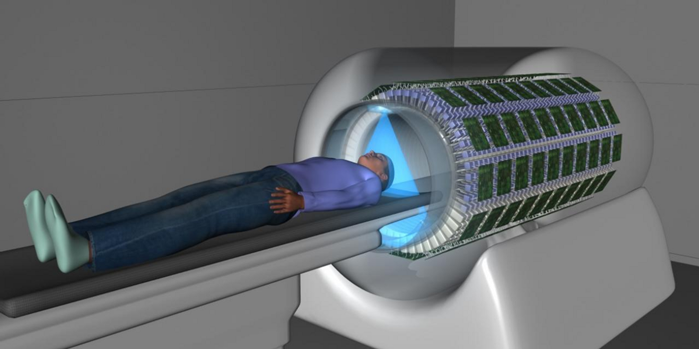
The conceptual drawing of the EXPLORER scanner (UC Davis/Biomedical Engineering)
Non-invasive diagnostic imaging like CT (computed tomography) and MRI (magnetic resonance imaging) allow doctors to perform proper diagnosis of diseases without puncturing the mucosa or skin, or reaching inside an internal body cavity. When used in conjunction with nuclear medicine modalities such as single-photon emission tomography (SPET) and positron emission tomography (PET), they can produce co-registered images that reveal not only anatomical and functional details of a body, but also quantitative information of disease-related biomarkers.
Last December, the US Food and Drug Administration approved a brand new PET/CT scanner called the EXPLORER. Co-developed by a research group at the University of California at Davis, led by professors Simon Cherry and Ramsey Badawi, it is the world’s first whole-body scanner.
Working with collaborators from other institutes including Zhongshan Hospital (Shanghai, China) and United Imaging Healthcare (Houston, TX), the team of biomedical engineers started their journey with a mandate to tackle the efficiency issue of conventional PET scanners.
Equipped with a ring-like detector approximately 10 inches, these scanners can only image a small region of the body that is at the center of the ring. To acquire a full-body scan, technicians would need to move the patient back and forth through the ring, which prolongs the window for a patient to stay on the scanner bed. (for example, an average of 20 minutes is required for a whole body PET scan.)
PET scans required the injection of positron-emitting radioactive probes, which rapidly decay during the imaging process. Therefore, the amount of radiation exposure becomes proportional to the size of the bodily region required for imaging: the bigger the area, the higher the exposure.
To resolve the problems, the UC Davis team combined 8 detector rings to form an 80 inches-long tube. With numerous improvement and tireless finetuning, the resulted PET camera can a whole-body scan using only 1/40th the radiation dose, or in 40 times faster, or with 40-fold stronger signal intensity and higher resolution.
The ground-breaking design has received world-wide recognition since its initial inception. Physics World, the flagship magazine of UK's Institute of Physics, praised the EXPLORER PET/CT as one of the top five breakthroughs of the year 2018.
The first clinical deployment of this brand new scanner is expected to happen this July.
Cardiac motion-capture using the EXPLORER Total Body PET scanner (Ramsey Badawi)
Source: Nature